
Advanced Technology
Precision Dentistry
When you seek care at our office, you are assured that Dr. Gordon and his staff utilize the latest in technology to enhance the quality and fit for your dental care.
Our practice uses high power magnification loupes to enhance the level of diagnosis and to deliver precise dental care to our patients. Operating telescopes (or as we refer to as “loupes”) are built into our glasses so we may provide you with the excellent dentistry you deserve. Dentistry is micro-surgery. You just can’t fulfill that level of care with the naked eye.
More Advanced Technology
Digital Imaging
Dr. Gordon carefully determines which and when radiographs are taken. There are many guidelines that we follow. Radiographs allow us to see everything we cannot see with our own eyes. Radiographs enable us to detect cavities in between your teeth, determine bone level, and analyze the health of your bone. We can also examine the roots and nerves of teeth, diagnose lesions such as cysts or tumors, as well as assess damage when trauma occurs.
Dental radiographs are invaluable aids in diagnosing, treating, and maintaining dental health. Exposure time for dental radiographs is extremely minimal. Dr. Gordon utilizes Digital Imaging Technologies within the office. With digital imaging, exposure time is about 60 – 80 percent less when compared to traditional radiographs. Digital imaging can also help us retrieve valuable diagnostic information. We may be able to see cavities better.
Digital imaging allows an enlarged image of your teeth and surrounding structures to be shown on the computer screen. This allows for easier transference of information and education to you the patient.
Digital imaging allows us to store patient images, and enables us to quickly and easily transfer them to specialists or insurance companies.
Intraoral Camera
Many patients, especially younger patients, are very familiar with the latest technology and are comfortable with this type of high tech equipment. Computers and TV screens are their primary method of information processing.
Dr. Gordon utilizes intraoral camera technology that helps enhance your understanding of his diagnosis. An intraoral camera is a very small camera – in some cases, just a few millimeters long. An intraoral camera allows our practice to view clear, precise images of your mouth, teeth and gums, in order for us to accurately formulate a diagnosis. With clear, defined, enlarged images, you see details that may be missed by standard mirror examinations. This can mean faster diagnosis with less chair-time for you!
Intra oral cameras also enable our practice to save your images in our office computer to provide a permanent record of treatments. These images can be printed for you, other specialists, and your lab or insurance companies.
Diagnodent
The Diagnodent aids in the detection of caries (decay) on the occlusal surface (biting surface) of a tooth. The Diagnodent uses laser technology to detect and quantify hidden or sub-surface caries by measuring fluorescence within the tooth structure. Even very small lesions are detected in the earliest stage, enabling Dr. Gordon to protect and preserve the tooth substance. The Diagnodent laser caries detection aid removes the doubt from treatment decisions regarding hidden caries or questionable stained grooves. The device’s ability to see the occlusal pits and fissures enables Dr. Gordon to treat sub-surface carious lesions with confidence.
VELscope
A New Tool for Oral Examinations
VELscope is a revolutionary hand-held device that provides dentists and hygienists with an easy-to-use adjunctive mucosal examination system for the detection of abnormal tissue.
Did you know?
- Oral Cancer kills one person every hour, 24 hours a day in North America
- The incidence of oral cancer has exceeded the incidence of cervical cancer
- The overall 5-year survival rate for oral cancer is 52%, but when discovered early, it increases to 80%-90%
- Pre-malignant changes actually start below the surface, at the basement membrane. The changes may not be apparent to the naked eye until the disease progresses to the surface
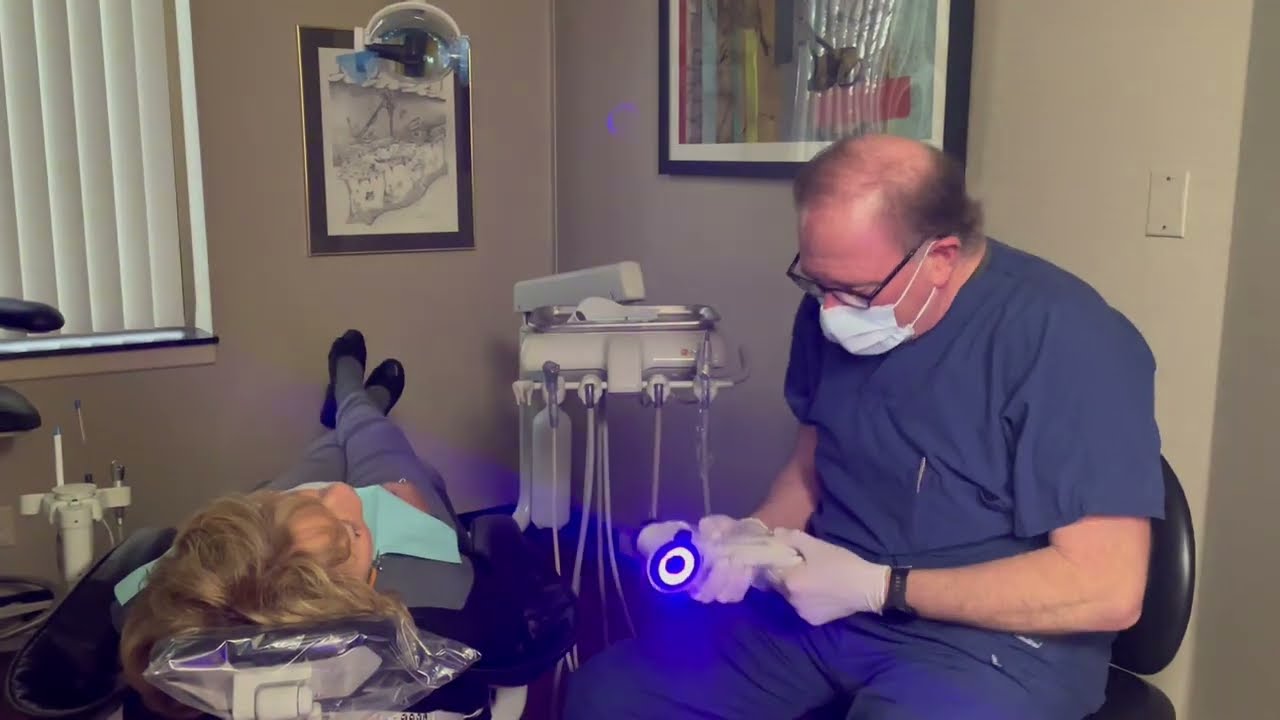
The VELscope emits a safe blue light into the oral cavity, which excites the tissue from the surface of the epithelium through the entire tissue layer to the basement membrane causing normal tissue to fluoresce. The clinician is then able to immediately view the different fluorescent responses not visible to the naked eye, helping differentiate between normal and abnormal tissue. In fact, VELscope is the only non-invasive adjunctive device clinically proven to help discover oral disease.
Dr. Gordon and his team are able to provide you with this technology at your First Visit and at your check-up exams, typically on an annual basis. How many little sores do we find in our mouths? Most of the time we simply ignore them and they go away. However, sometimes our mouths keep “secrets”. That’s why we are proud to offer the VELscope Enhanced Assessment System, a potentially life-saving technology, to our patients.
The VELscope helps:
- Improve our assessment of your overall oral health.
- Ensure that the delicate tissues of your mouth are healthy.
- Protect you from oral disease, including oral cancer.
All this in two minutes, with no rinses, stains or discomfort.